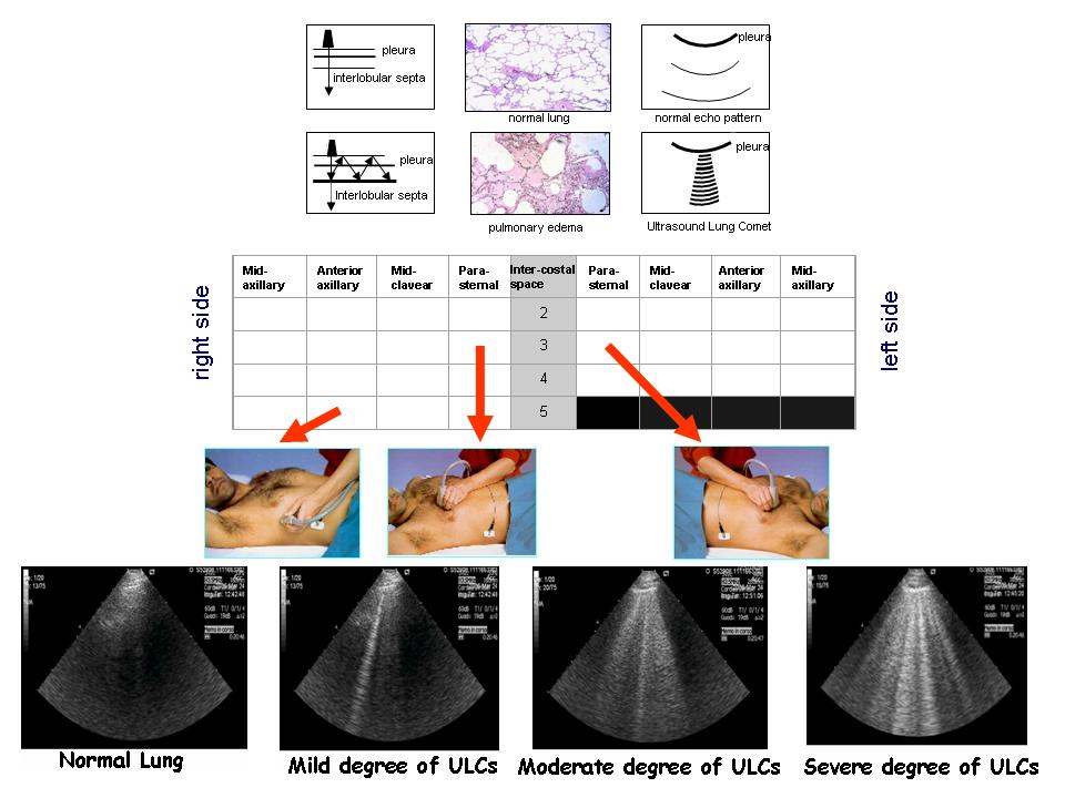
Ultrasound Lung Comets are a novel chest echographic sign of subpleural septa of increased thickness, due to reversibile water accumulation (as it happens in cardiogenic pulmonary edema) and/or irreversible fibrosis tissue in lung disease (as it happens in the interstitial syndrome of pulmonary fibrosis). In heart failure patients, the presence, site and number of ultrasound lung comets allows detection, location and quantification of extra-vascular lung water. With chest sonography, the normal lung is "black", moderately diseased lung (with interstitial water) is "black and white" (with white lines corresponding to comets) and markedly diseased lung (with alveolar edema) is "white" (diffusely bright). The potential main domain of application of ultrasound lung comets in cardiology is the heart failure patient. The practical appeal of the technique stems from its "green" (non-ionizing, without biological hazard for the patients and the operator) and "light" (portable, low cost, basic technological requirements, elementary skills) nature.
Immagini:

