As a result of their importance and value, works of art require the development of non-destructive and non-contact techniques to the greatest extent possible. Moreover, because of the immovable nature of some artworks (mural paintings, sculptures, buildings, etc.), the development of portable instrumentation for in-situ investigation, analysis and monitoring is an important requirement. To date, only some non-contact portable equipments are available in a number that appears low compared to the large variety of conventional laboratory instrumentations. Realizing these shortcomings, our main goal is to develop new portable techniques to study the surface molecular composition of art works.
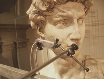
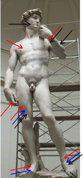
As example of this research line, we report recent results obtained on developing mid FT-IR reflectance spectroscopy to non-destructively investigate the alteration state of marble surfaces by means of portable instrumentation. Following an accurate laboratory setting procedure of the methodology, the technique was successfully applied within a wide project to study the conservation state of the Michelangelo's David led by the CNR Institute ICVBC. In-situ non-destructive measurements were carried out by a portable FT-IR equipment located on scaffolding arranged in order to reach the probed areas (Figure 1). During measurement acquisitions a mechanical arm or hand positioning of the fiber optic probe have been used as required by the morphology of the surface.The investigation was focused on marble visible alteration such us gray incrustations, residual black crusts, yellowish or dark patinas. By analyzing the collected spectra three different kinds of contaminations were identified. First, sulphates were clearly detected on residual black crust (right hand forefinger) as well as on gray incrustation (for instance, on the right foot and the right chest). Traces of sulphates have been found also in areas that are not macroscopically altered. Thus, sulphate contamination greater than a few micromole/cm2 was to be spread all over the statue, with higher concentrations on specific zones (Figure 2, red arrows). Second, oxalates were usually identified on yellowish patinas, as on the left foot. Unlike the sulphates, oxalate contamination resulted to be mainly localized in the lower part of the statue (Figure 2, blue arrows). Third, spectral features of organic components (mainly C=O and C-H stretchings) were observed in some spectra; when they appeared well resolved from the substrate absorption an identification of the organic component was attempted (Figure 2, black arrows). Following the results obtained from laboratory measurements on reference standards, the organic component was identified as wax.
Vedi anche:
Immagini:


