The cerebral blood flow is finely regulated by neuronal activity. When neurons in a specific brain region are highly activated following a distinct set of stimuli - for example, the neurons in the auditory cortex during listening to a concert - the blood flow in that region increases in a temporally and spatially coordinated manner. This phenomenon is called functional hyperemia and is a fundamental event in brain function. It was first described by the italian Angelo Mosso and later by the british Charles Sherrington more than a century ago. The molecules, signaling pathways and the ultimate mechanism at the basis of this phenomenon were largely undefined despite the intense investigation that was performed during the last decade and the increasing use of this phenomenon in basic and clinical neurosciences, as revealed by the increasing importance of modern brain imaging techniques.
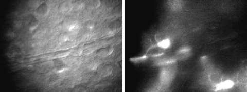
In our study, we show that a local activation of neurons - that triggers the dilation of cerebral arterioles - also triggers intracellular calcium elevations in astrocytes. We were able to show that these calcium responses spread intracellularly from astrocyte processes in contact with the synapse to the endfeet in contat with the arterioles. The inhibition of these responses in glial cells results in the impairment of neuronal activity-dependent vasodilation, both in brain slices and in in vivo experiments, while the selective activation by a patch pipette of single astrocytes in contact with arterioles triggers their relaxation.
Astrocyte-mediated control of arterioles appears to be mediated by a cyclooxygenase product, possibly the powerful vasodilator prostaglandin E2.
Information on the status of neuronal activity can, therefore, be encoded by astrocytes in a defined frequency of oscillations and transferred to astrocyte endfeet in contact with blood vessels. There, [Ca2+]i oscillations can be translated into a pulsatile release of vasoactive agents that result in the dilation of cerebral arterioles. The specific activation of this neuron-to-astrocyte signalling appears, therefore, a crucial step in the mechanism that governs the phenomenon of functional hyperemia.
Our results provide a mechanistic understanding of the basis for the neuronal control of blood flow and thus the molecular basis on which modern brain imaging techniques, such as positron emission tomography (PET) and functional magnetic resonance imaging (fMRI), rely.
The astrocyte role in the control of cerebral blood flow may represent a clue for understanding the mechanism underlying cerebrovascular deficiencies occurring in numerous central nervous system pathologies such as Alzheimer and Parkinson diseases. The underlying hypothesis is that a damage to the astrocytes may result in the failure of these cells to respond properly to neuronal signals and, in turn, to order the dilation of blood vessels. Under these conditions, neurons can not be provided with the necessary energetic substrates. The ultimate result of this process can be an in initial impairement of specific neuronal properties, such as memory function, that can be followed by cell death. Astrocytes may, therefore, represent a potential target for a better understanding of the cellular mechanism at the basis of neurodegenerative diseases of the central nervous system as well as for the development of novel therapeutic approaches for these diseases.
Vedi anche:
Immagini:

